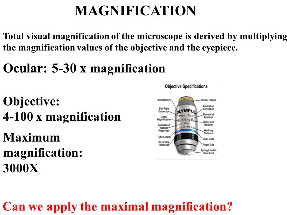
Laser Scanning Microscope Maximum Magnification
Micro Small Scopeo To Watch Microscopy The Relative Sizes Of Molecules Cells And Organisms Ppt Download

What Is Confocal Laser Scanning Microscopy
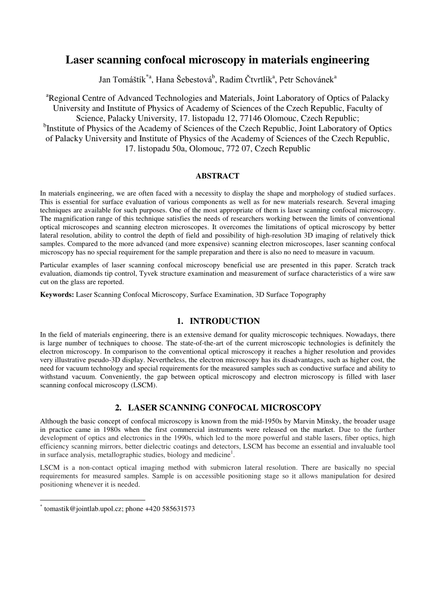
Pdf Laser Scanning Confocal Microscopy In Materials Engineering

Confocal Laser Scanning Microscopy An Overview Sciencedirect Topics

Imaging Surface Characterisation Australian National Fabrication Facility Queensland Node

Confocal Laser Scanning Microscope Labcompare Com
Relatively thick specimens can be imaged in successive volumes by acquiring a series of sections along the optical z axis of the microscope.
Laser scanning microscope maximum magnification. This means that we can view visual sections of tiny structures that. Capturing multiple two dimensional images at different depths in a sample enables the. Confocal laser scanning fluorescence microscope. A transmission electron microscope tem produces a 2d image of a thin sample and has a maximum resolution of 500000.
Radiation in a lm. With confocal laser scanning microscopy clsm we can find out even more. Clsm combines high resolution optical imaging with depth selectivity which allows us to do optical sectioning. A confocal laser scanning microscope or also known as laser scanning confocal microscope is used to obtain high resolution images and generate 3d reconstructions through direct optical sectioning to provide clear images from a range of depths of thicker specimens for nanometer level imaging and measurement.
The maximum magnification of a light microscope. The confocal laser scanning microscope clsm is a microscope which focuses only on a single focal plane and the unfocused plane will not be visualized. Additional advantages of scanning confocal microscopy include the ability to adjust magnification electronically by varying the area scanned by the laser without having to change objectives. Light microscope laser scanning confocal microscope transmission electron microscope tem and scanning electron microscope sem light.
Fluorescent microscopy not only makes our images look good it also allows us to gain a better understanding of cells structures and tissue. In the past the traditional laser microscope excited the whole thickness of the sample resulting in saturated blurry images and sometimes visualizing false colocalization images. Lets you look specifically at parts of a cell such as individual proteins by labelling them with fluorescence. Lens used in a lm.
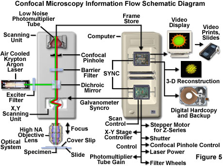
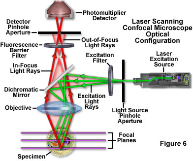
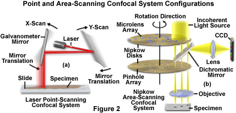
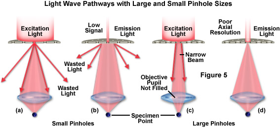
This feature is termed the zoom factor and is usually employed to adjust the image spatial resolution by altering the scanning laser sampling period. Laser scanning confocal microscopy laser scanning confocal microscopes employ a pair of pinhole apertures to limit the specimen focal plane to a confined volume approximately a micron in size. Confocal microscopy most frequently confocal laser scanning microscopy clsm or laser confocal scanning microscopy lcsm is an optical imaging technique for increasing optical resolution and contrast of a micrograph by means of using a spatial pinhole to block out of focus light in image formation. Lscm laser scanning confocal microscope parameter value comment maximum ip pixel resolution 1270 x 1000 pixel user adjustable maximum field of view 1 ccd equivalent 9 6 x 12 8mm.
Lets you look at thin slices in a sample while keeping sample intact.
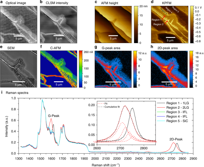
Confocal Laser Scanning Microscopy For Rapid Optical Characterization Of Graphene Communications Physics
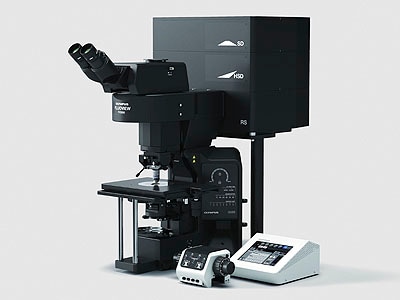
Zeiss Lsm 880 W Fast Airyscan Bacterial Imaging Cluster

Eye Of A Blue Dragonfly By Igor Siwanowicz Confocal Laser Scanning Microscopy Symmetry Microscopic Photography Microscopic Images Micro Photography

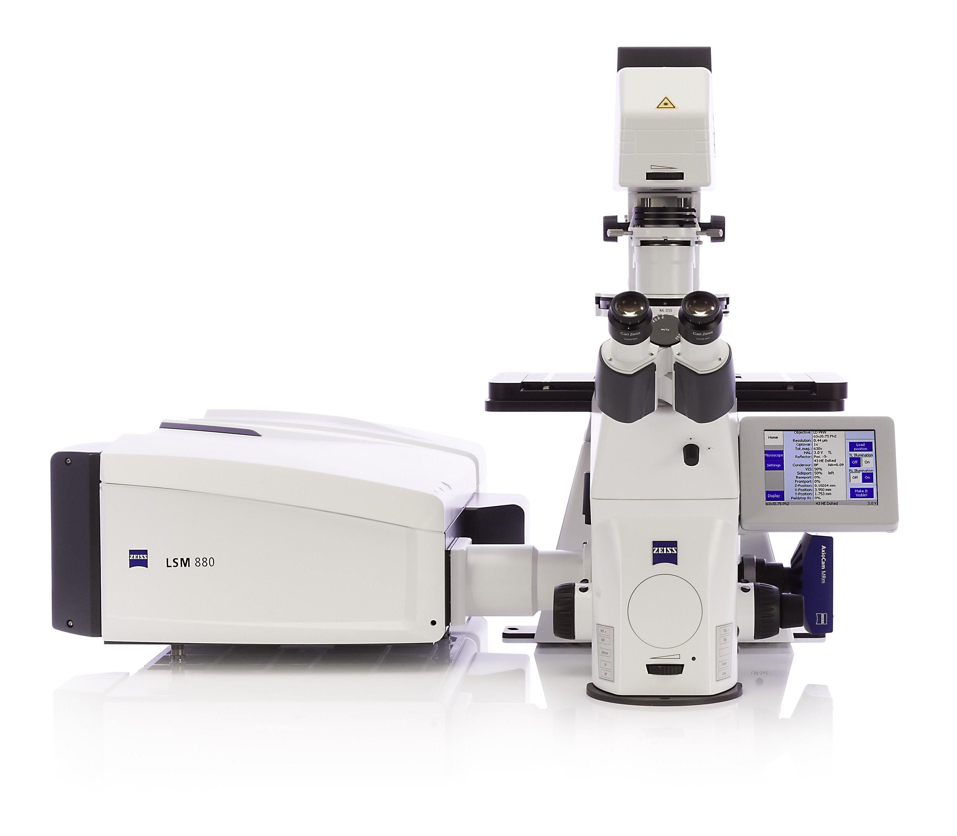
Inverted Zeiss Lsm880 Laser Scanning Confocal Microscope With Airyscan Cell Sciences Imaging Facility Csif
Confocal Microscopy Introduction Olympus Life Science
Fv3000 Confocal Laser Scanning Microscope From Olympus Life Science Solutions Get Quote Rfq Price Or Buy
Zeiss Microscopy Online Campus Live Cell Imaging Microscopy Techniques

Laser Scanning Microscope 13 Steps With Pictures Instructables

Overview Of Confocal Laser Scanning Microscopes

Pin By Joe Mykietyn On Insects Nikon Small World Microscopic Photography Small World

New Microscope Shows The Quantum World In Crazy Detail Quantum World Electron Microscope Properties Of Materials

Appendages Of A Common Brine Shrimp Dr Igor Siwanowicz Howard Hughes Medical Institute Hhmi Confoc Nikon Small World Brine Shrimp Microscopic Photography

Laser Scanning Microscopes Keyence America

Advantages And Limitations Of Scanning Electron Microscopy Sem And Download Table

Eyes Of A Harvestman Aka Daddy Longlegs Spider Photo By Photographer Igor Siwanowicz La Photographie Microscopique Galeries De Photos Jolie Photo
Confocal Microscopy Confocal Microscope Scanning Systems Olympus Life Science

Confocal Laser Scanning Microscope An Overview Sciencedirect Topics

Optical Microscope All Industrial Manufacturers Videos
Https Encrypted Tbn0 Gstatic Com Images Q Tbn 3aand9gcr3fysoxor5w4y0kayjtt5nby84 Yhi3vdxn3rx2 E Usqp Cau
Olympus Launches Two Types Of Upright Fv3000 Confocal Laser Scanning Microscope 2017 News Olympus
Zeiss Microscopy Online Campus Digital Imaging Considerations

Diatom Coloured Scanning Electron Micrograph Sem Of A Triceratium Sp Diatom Biologia Taide

Use Of Confocal Laser Scanning Microscopy For Biofilm Investigation On Paints Under Field Conditions Sciencedirect

This Is A Scanning Electron Micrograph Of A House Fly S Eye Microscopic Images Microscopic Photography Microscopic

Confocal Laser Scanning Microscopy And Scanning Electron Microscopy Of Tissue Ti Implant Interfaces Sciencedirect

See The Tiny Carnivorous Monster That Just Won Olympus S Micro Photo Prize Microscopic Photography Patterns In Nature Microscopic
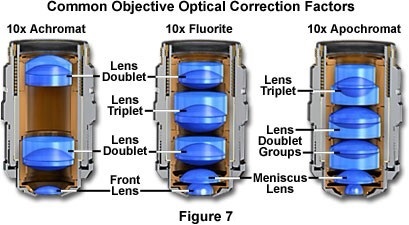
Confocal Microscopy Confocal Microscope Objectives Olympus Life Science

Eye Of A Damselfly Image By Igor Siwanowicz Max Planck Institute For Neurobiology Munich Germany Microscopic Photography Micro Photography

Microscopy And Image Analysis Mcnamara 2017 Current Protocols In Human Genetics Wiley Online Library

Visual Surround System By Blepharopsis Laser Scanning Confocal Microscope Image Of Zebra Jumping Spider S Dinosaur Bones Jumping Spider Sorel Winter Boot

Award Winning Images Of Really Tiny Things On Earth Microscopic Photography Scanning Electron Microscope Microscopic Images

Aloe Vera Under The Light Microscope Microscopic Photography Under The Lights Microscopic
.jpg?rev=33A9)
Fv1200 Olympus Life Science

Olympus News Release Confocal Laser Scanning Microscope Ols 3000 World S Highest Resolution Of 0 12µm

Confocal Laser Scanning Microscope Specification Price Image Bio Equip In China

Laser Scanning Confocal Microscopy Java Tutorial Olympus Life Science

Inverted Zeiss Lsm 780 Multiphoton Laser Scanning Confocal Microscope Cell Sciences Imaging Facility Csif

Alimentos Bajo El Microscopio Coliflor Microscopic Food Animals Macro And Micro

Zeiss Microscopy Online Campus Introduction To Spinning Disk Microscopy

Mice Are The Most Commonly Used Mammalian Research Model With Hundreds Of Established Inbred Outbred And Transgenic Strains They Are Microscopic Photography Science Illustration Tiny World

Tiny Grandeur Stunning Photos Of The Very Small Nikon Small World Small World Microscopic Photography

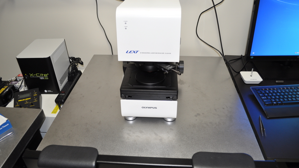
Olympus News Release Olympus Launches Lext Ols3100 Confocal Laser Scanning Microscope