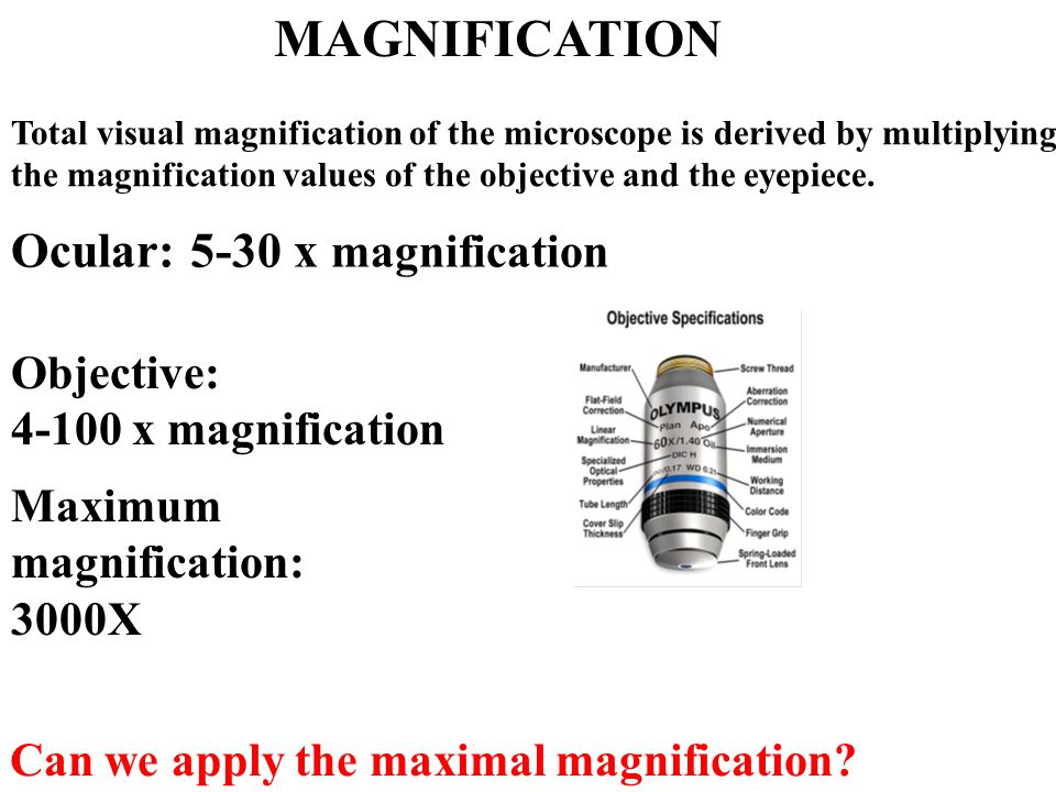
Laser Scanning Microscope Max Magnification

What Is Confocal Laser Scanning Microscopy

Confocal Laser Scanning Microscopy An Overview Sciencedirect Topics
Confocal Microscopy Introduction Olympus Life Science
Confocal Microscopy Confocal Microscope Scanning Systems Olympus Life Science
Fv3000 Confocal Laser Scanning Microscope From Olympus Life Science Solutions Get Quote Rfq Price Or Buy

Inverted Zeiss Lsm880 Laser Scanning Confocal Microscope With Airyscan Cell Sciences Imaging Facility Csif
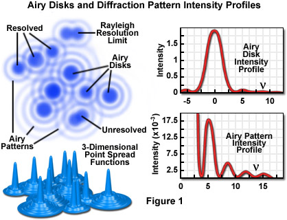
This means that we can view visual sections of tiny structures that.
Laser scanning microscope max magnification. Additional advantages of scanning confocal microscopy include the ability to adjust magnification electronically by varying the area scanned by the laser without having to change objectives. Separating light waves with lasers and with. Capturing multiple two dimensional images at different depths in a sample enables the. With confocal laser scanning microscopy clsm we can find out even more.
Relatively thick specimens can be imaged in successive volumes by acquiring a series of sections along the optical z axis of the microscope. The confocal laser scanning microscopes enable researchers to create detailed 3d pictures of cell organelles and to examine live cells through incubation systems that facilitate the study of cellular changes over time. This feature is termed the zoom factor and is usually employed to adjust the image spatial resolution by altering the scanning laser sampling period. In some cases specimens should be sampled at more than 2 3 times the highest information frequency to allow for the possibility that the highest frequency was misjudged.
Confocal microscopy most frequently confocal laser scanning microscopy clsm or laser confocal scanning microscopy lcsm is an optical imaging technique for increasing optical resolution and contrast of a micrograph by means of using a spatial pinhole to block out of focus light in image formation. The laser scans across the object and an image is built up pixel by pixel on a screen. A confocal laser scanning microscope or also known as laser scanning confocal microscope is used to obtain high resolution images and generate 3d reconstructions through direct optical sectioning to provide clear images from a range of depths of thicker specimens for nanometer level imaging and measurement. A scanning electron microscope sem produces a 3d image of a sample by bouncing electons off and dectecting them at multiple detectors.
In the confocal laser scanning microscope the highest frequency to be sampled f is imposed by the optical system and for a particular resolution specification. Magnification image contrast laser intensity detector sensitivity flip rotate image line scan and area scan modes are user selectable includes laser scanning confocal microscope lscm controller controller cables galvanometer data communication power interlock and connector kit. A transmission electron microscope tem produces a 2d image of a thin sample and has a maximum resolution of 500000. The confocal laser scanning microscope s aim was not to further increase magnification but to make clearer.
It has a maximum magnification of about 100000. Pongali raghavendra thammineni pullaiah in advances in cell and molecular diagnostics 2018.

Confocal Laser Scanning Microscope Labcompare Com
Confocal Microscopy Resolution And Contrast In Confocal Microscopy Olympus Life Science
Micro Small Scopeo To Watch Microscopy The Relative Sizes Of Molecules Cells And Organisms Ppt Download

A Practical Guide For Fluorescent Confocal Microscopy The Marder Lab

Zeiss Lsm 900 Confocal Laser Scanning Microscope From Carl Zeiss Microscopy Biocompare Com

Laser Scanning Microscope 13 Steps With Pictures Instructables

Overview Of Confocal Laser Scanning Microscopes

Advantages And Limitations Of Scanning Electron Microscopy Sem And Download Table

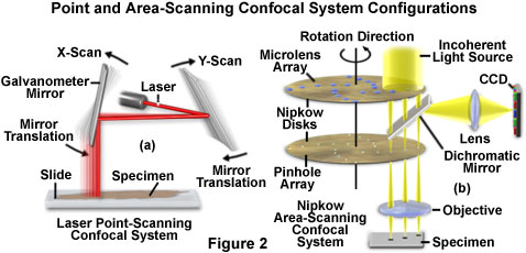
Zeiss Microscopy Online Campus Introduction To Spinning Disk Microscopy

Olympus News Release Confocal Laser Scanning Microscope Ols 3000 World S Highest Resolution Of 0 12µm

Confocal Laser Scanning Microscope An Overview Sciencedirect Topics

33 Laser Scanning Confocal Microscopy And Laser Microdissection Musculoskeletal Key

Laser Scanning Microscopes Keyence America

Rp Photonics Encyclopedia Optical Profilometers Non Contact Optical Surface Profile Measurements Interferometer Coherence Oct Focus Variation Digital Holography Triangulation Time Of Flight Structured Light Performance Factors Geometric
Olympus Launches Two Types Of Upright Fv3000 Confocal Laser Scanning Microscope 2017 News Olympus

Scanning Optical Microscopy Emmi Laser Scan Obirch Som

Laser Scanning An Overview Sciencedirect Topics
Confocal Microscopy Confocal Microscope Objectives Olympus Life Science
Https Encrypted Tbn0 Gstatic Com Images Q Tbn 3aand9gcr3fysoxor5w4y0kayjtt5nby84 Yhi3vdxn3rx2 E Usqp Cau

Adaptive Optics For Biomedical Microscopy Optics Photonics News
.jpg?rev=33A9)
Fv1200 Olympus Life Science

Inverted Zeiss Lsm 780 Multiphoton Laser Scanning Confocal Microscope Cell Sciences Imaging Facility Csif

Imaging Surface Characterisation Australian National Fabrication Facility Queensland Node

Confocal Laser Scanning Microscope Specification Price Image Bio Equip In China

Confocal Laser Scanning Microscopy And Scanning Electron Microscopy Of Tissue Ti Implant Interfaces Sciencedirect

Olympus News Release Olympus Launches Lext Ols3100 Confocal Laser Scanning Microscope
Pdf Optimal Lens Design And Use In Laser Scanning Microscopy
Confocal Microscopy Signal To Noise Considerations Olympus Life Science
Confocal Microscopy Colocalization Of Fluorophores In Confocal Microscopy Olympus Life Science

Pdf Application Of Confocal Laser Scanning Microscopy In Dentistry
Pdf Surface Roughness Determination Using Laser Scanning Confocal Microscope Zeiss Lsm 700

3d Laser Scanning Microscopy Model Vk X200 Keyence Lnnano Lnnano
Methods To Probe The Formation Of Biofilms Applications In Foods And Related Surfaces Analytical Methods Rsc Publishing Doi 10 1039 C9ay02214g
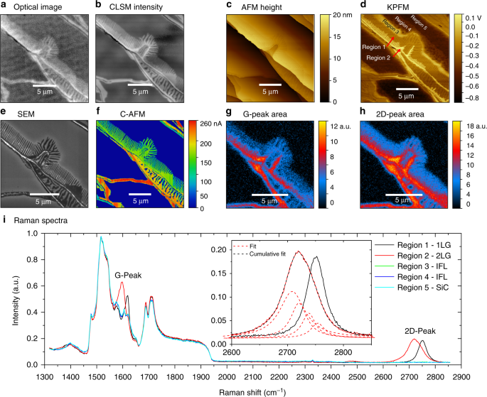
Confocal Laser Scanning Microscopy For Rapid Optical Characterization Of Graphene Communications Physics

Laser Scanning Confocal Microscopy Java Tutorial Olympus Life Science

Biophotonics Lecture 16 November Fourier Plane Point Object Image F F F F Magnification M 1 Angles Sin Sin Magnification Ppt Download
Pdf Any Way You Slice It A Comparison Of Confocal Microscopy Techniques

Pdf A 3d Imaging And Visualization Workflow Using Confocal Microscopy And Advanced Image Processing For Brachyuran Crab Larvae

Pdf Fluorescence Microscopy

Laser Raman Microscopy Ramantouch Ramanforce Nanophoton Corp

Smart Control For Resonant Galvo Scanners Learn Share Leica Microsystems

