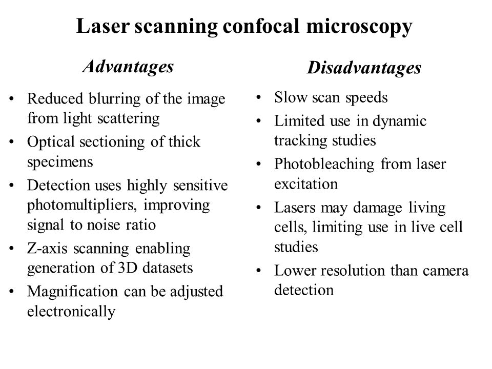
Laser Scanning Microscope Disadvantages

What Are The Limitations Of Confocal Laser Scanning Microscopes Quora

Near Field Scanning Optical Microscopy Advantages And Disadvantages
Confocal Laser Scanning Microscopy Clsm

Modern Laser Scanning Confocal Microscopy Bayguinov 2018 Current Protocols In Cytometry Wiley Online Library

The Use Of Laser Scanning Confocal Microscopy Lscm In Materials Science Hovis 2010 Journal Of Microscopy Wiley Online Library

Manual Capsulorhexes Above And Catalys Capsulotomies Below Stained With Trypan Blue Catalys Capsulotomies Exhibit Precisio Cataract Surgery Cataract Laser

Light microscope transmission electron microscope scanning electron microscope and laser scanning confocal microscope.
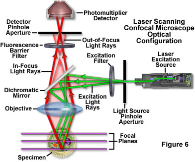
Laser scanning microscope disadvantages. When investigating multilayer structures the true surface of a substrate can be observed through a surface coating. In contrast conventional widefield microscopes use mercury or xenon based arc discharge lamps to provide a full range of excitation wavelengths in the ultraviolet visible and near infrared spectral regions. Laser scanning confocal fluorescence microscopy. A typical confocal uses raster scanning which means it scans the specimen point by point.
The laser scans across the object and an image is built up pixel by pixel on a screen. Electrons pass through the specimen and form an image. Imaging frame rates are typically slower for single point laser. A thick section of fluorescently stained human medulla in widefield fluorescence exhibits a large amount of glare from fluorescent structures above and below the focal plane figure 1 a.
The confocal laser scanning microscope s aim was not to further increase magnification but to make clearer. Comparing to a wide field detection taking a snapshot of the whole field of view it is quite slow. The primary advantage of laser scanning confocal microscopy is to produce thin optical sections through fluorescent specimens that have a thickness beyond 50 micrometers. Confocal laser scanning microscopes can have a programmable sampling density and very high resolutions while nipkow and pam use a fixed sampling density defined by the camera s resolution.
Of course you can make it faster by compromising sensitivity resolution etc and there are some special implementation of confocal such as spinning disk confocal to resolve this issue. The thickness of the coating can be determined by observing the 2 peaks in the axial intensity variation. Laser scanning confocal microscopy. Advantages of confocal laser scanning microscopy industrial applications of confocal microscopy thin film profiling.
Electrons are reflected off the specimen to produce a 3d image. A the use of microscopy to observe and investigate different types of cell and cell structure in a range of eukaryotic organisms to include an appreciation of the images produced by a range of microscopes. Black and white images. When imaged with a laser scanning confocal microscope figure 1 d the.
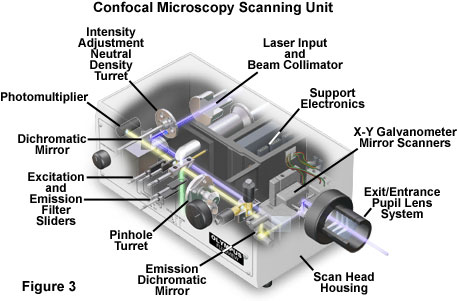
Spinning disk systems provide an alternative means of obtaining a full frame high speed confocal image in real time.
Zeiss Microscopy Online Campus Live Cell Imaging Microscopy Techniques

World S First White Lasers Demonstrated More Luminous Energy Efficient Than Leds White Lasers Look To Be The Future In Lighting And Li Fi Or Light Based Wir Futuristic Technology Nanotechnology Technology
Spinning Disk Vs Laser Scanning Confocal Microscopes Features Oct 2004 Photonics Spectra
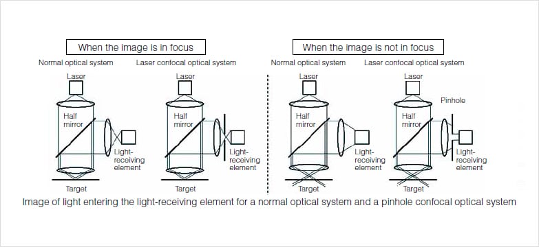
Confocal Microscopy Introduction Olympus Life Science
If You Need Any Optical And Ophthalmic Device Pls Contact Me Email Lisafan Hyvisionstar Com Eye Health Ophthalmic Equipment Optical
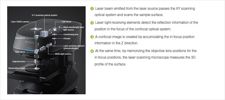
Profile Measuring Laser Microscopes Instruments Used For Roughness Measurements Introduction To Roughness Keyence America

Pdf Confocal Scanning Optical Microscopy And Its Applications For Biological Specimens Semantic Scholar

Tevo Tornado X Axis Tensioner Remix By Gabix Thingiverse Tornado Remix 3d Printing

Python Programming Logo In Stained Glass 105 00 Via Etsy This Would Be A Fun Thing To Put In One Of My Wind Python Programming Language Logo Learn To Code
Olympus Fluoview Resource Center Spectral Bleed Through Artifacts In Confocal Microscopy
.jpg)
The Benefits Of Using A Confocal Microscope

Confocal Microscopy An Overview Sciencedirect Topics

Orlas Station Machine Design Cnc Design Industrial Machine
Fluorescence And Confocal Microscopy Ppt Video Online Download
Confocal Laser Scanning Microscopy Springerlink

Tattooremovalproducts Laser Hair Removal Machine Hair Removal Machine Tattoo Removal

What Is Spinning Disk Confocal Microscopy

What Are The Main Differences Between An Sem An Esem An Sem Fib And An S Tem Horiba
Https Encrypted Tbn0 Gstatic Com Images Q Tbn 3aand9gctuan2diibxh Akqhct Gs3wlxe49owmw0 Ho3imvfwzh93skjl Usqp Cau
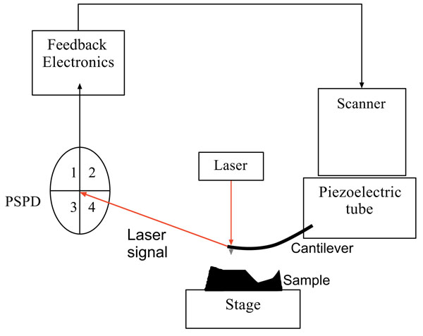
Review Of Progress In Atomic Force Microscopy Fulltext

Future Directions For Induced Pluripotent Stem Cells Research Stem Cell Research Stem Cells Cell

Fluorophores An Overview Sciencedirect Topics

Confocal Laser Scanning Microscopy An Overview Sciencedirect Topics

Femtosecond Laser Setups For Cell Membrane Poration

Confocal Laser Scanning Microscope An Overview Sciencedirect Topics

Pin On Eye Facts

Automotive Ecu Market Size Worth 60 5 Billion By 2025 Cagr 6 8 Grand View Research Inc Financial News Best Solar Panels Online Journal

Attendance In Excel Sheet Using Rfid Rc522 Hackster Io Arduino Rfid Arduino Beginner

Confocal Laser Scanning Microscopy An Overview Sciencedirect Topics

Starter Microscopes Which Image Is From The Light Microsope How Do You Know Ppt Download
Non Contact Surface Roughness Profile Measuring Instruments Instruments Used For Roughness Measurements Introduction To Roughness Keyence America
Http Www Microscopist Co Uk Wp Content Uploads 2017 04 Artefacts In Confocal Pdf
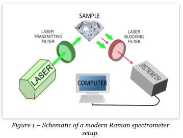
Advantages And Disadvantages Of Raman Fourier Transform Infrared Spectroscopy Ftir In The Gemological Field Agta

Pdf The Basics Of Confocal Microscopy
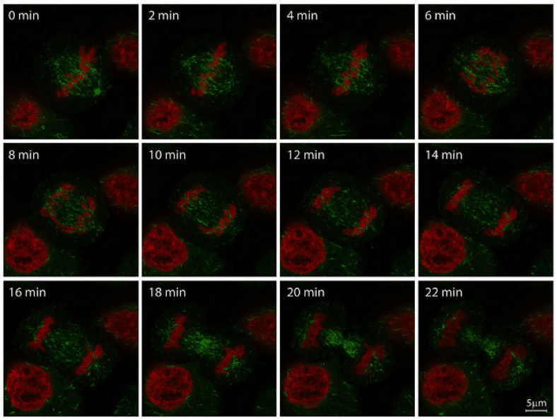
Tools For Shape Analysis Of Vascular Response Using Two Photon Laser Scanning Microscopy By Han Van Triest Committee Prof Dr Ir B M Ter Haar Romeny Ppt Download

Benefits Of Confocal Microscopy In Modern Life Science Applications Vision Blog

Direct Writing An Overview Sciencedirect Topics
Live Cell Imaging Choosing The Right Technique
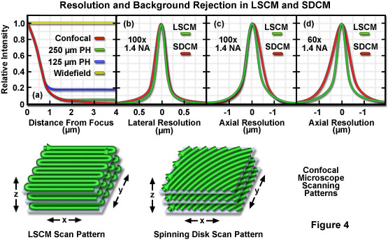
Zeiss Microscopy Online Campus Introduction To Spinning Disk Microscopy

Excimer Lasers Ento Key

Digital Podium Is The State Of The Art Solution For Smart Conference Room Smart Classroom And Auditorium I Interactive Presentation Digital Activities Podium
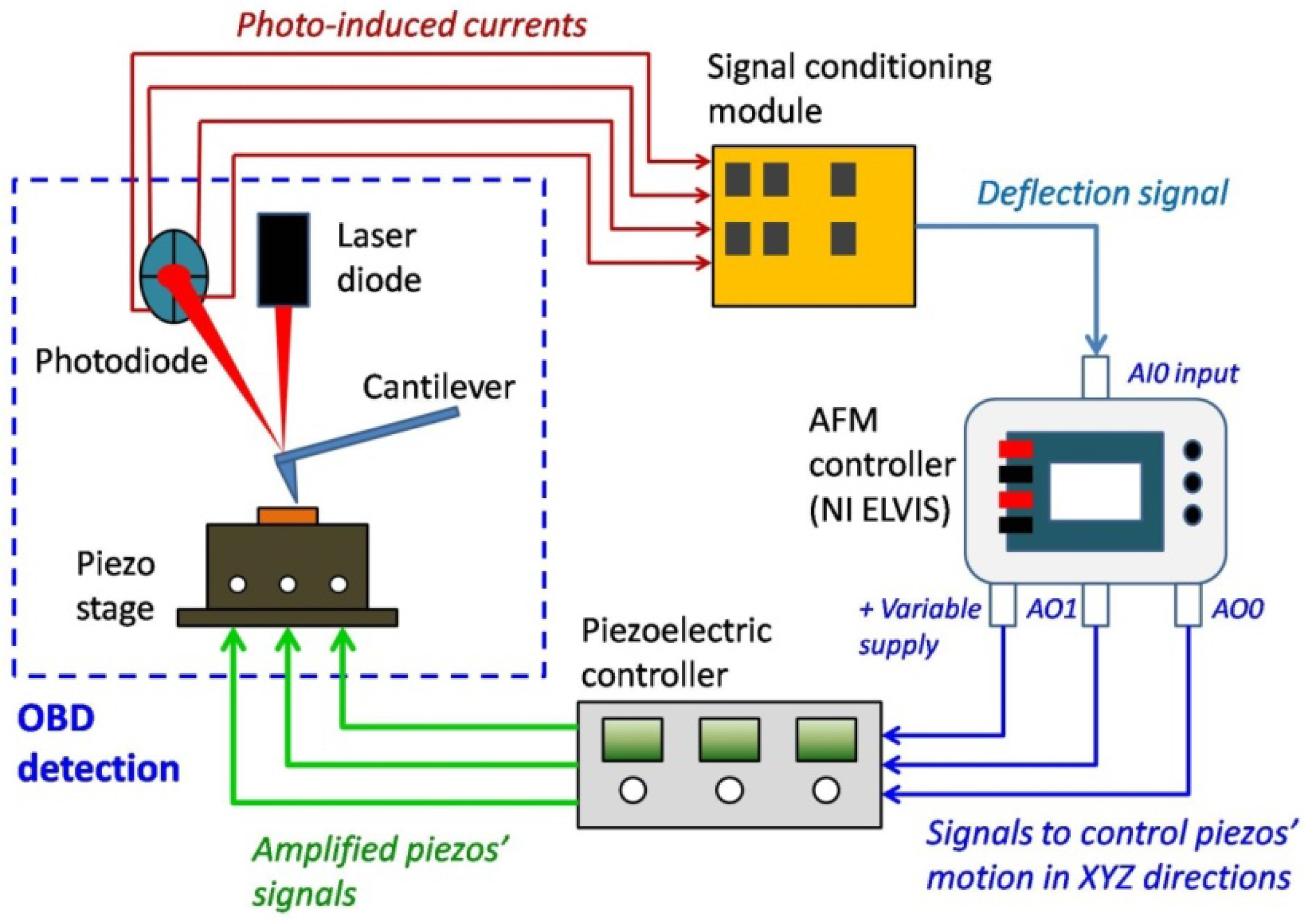
Applied Sciences Free Full Text Optical Beam Deflection Based Afm With Integrated Hardware And Software Platform For An Undergraduate Engineering Laboratory Html

