Laser Scanning Confocal Microscope Maximum Magnification

Another drawback is that duv microscopes do not have the capability for surface topography 3d imaging.
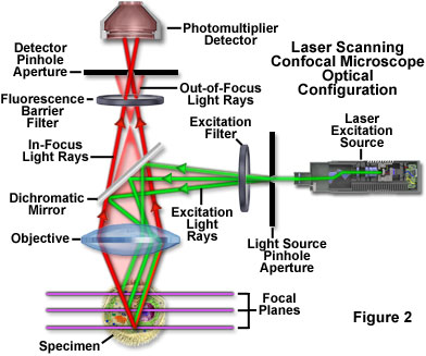
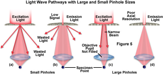
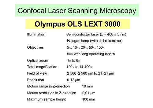
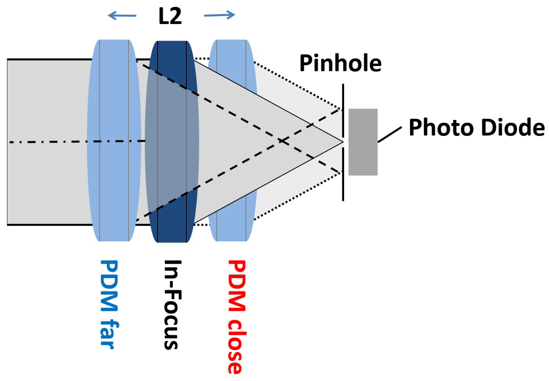
Laser scanning confocal microscope maximum magnification. The confocal laser scanning microscope s aim was not to further increase magnification but to make clearer. Laser scanning confocal microscopy laser scanning confocal microscopes employ a pair of pinhole apertures to limit the specimen focal plane to a confined volume approximately a micron in size. What is the maximum magnification of the scanning electron microscope. This resulted in a costly microscope system with magnification limited to 1000x.
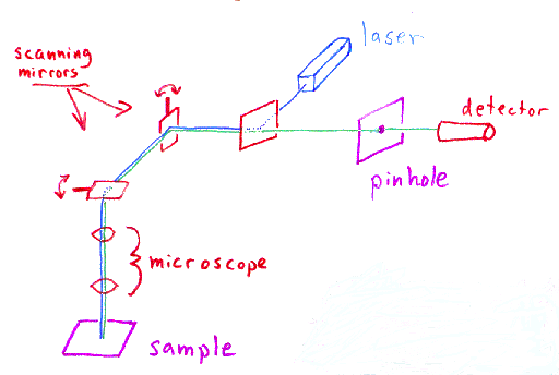
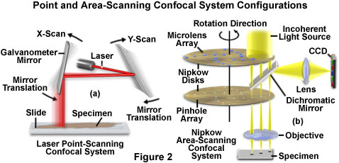
Relatively thick specimens can be imaged in successive volumes by acquiring a series of sections along the optical z axis of the microscope. The laser scanning microscope uses a scanning design called beam scanning where the laser image path is scanned in a raster pattern on the surface of the sample. Capturing multiple two dimensional images at different depths in a sample enables the. In the past the traditional laser microscope excited the whole thickness of the sample resulting in saturated blurry images and sometimes visualizing false colocalization images.
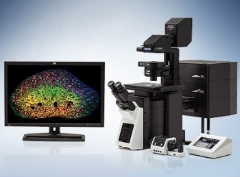
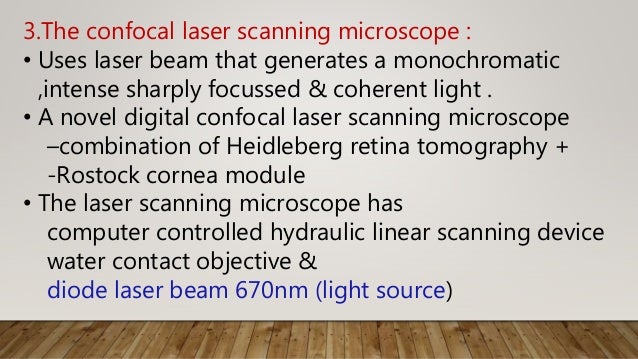
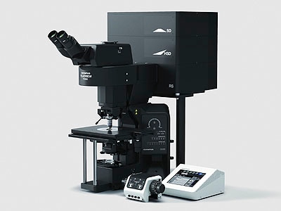
The scanning electron microscope has a magnification range from 15x to 200 000x reached in 25 steps and a resolution of 5. A confocal laser scanning microscope or also known as laser scanning confocal microscope is used to obtain high resolution images and generate 3d reconstructions through direct optical sectioning to provide clear images from a range of depths of thicker specimens for nanometer level imaging and measurement.















.jpg)


